

Digital Implant Planning vs. Traditional Methods
Quick Summary
- Diagnostic clarity: Digital implant planning pairs 3D CBCT scans with specialized software to map bone volume, nerve pathways, and tooth positions before any procedure begins.
- Prosthetic accuracy: Using a custom surgical guide reduces angular and positional deviation compared to unguided freehand placement.
- Predictable timelines: Pre-planning allows clinicians to determine if a temporary tooth is viable on the day of surgery, while standard healing typically spans 8 to 12 weeks.
- Transparent cost scope: Evaluating bone density in advance prevents unexpected surgical procedures mid-treatment, keeping single-tooth therapy within a clear €2,500 to €3,100 framework.
Missing a tooth impacts chewing stability, speech, and long-term oral balance. For years, dental implant placement relied primarily on two-dimensional imaging and the tactile instincts of the surgeon on the day of surgery.
Today, modern implantology approaches replacement teeth through restorative-driven digital engineering. Planning the final crown first ensures the underlying titanium fixture is placed in the exact biological and mechanical position required for lasting function.
What Exactly Is Digital Implant Planning? (And Why “3D-Guided” Is No Longer Optional in 2026)
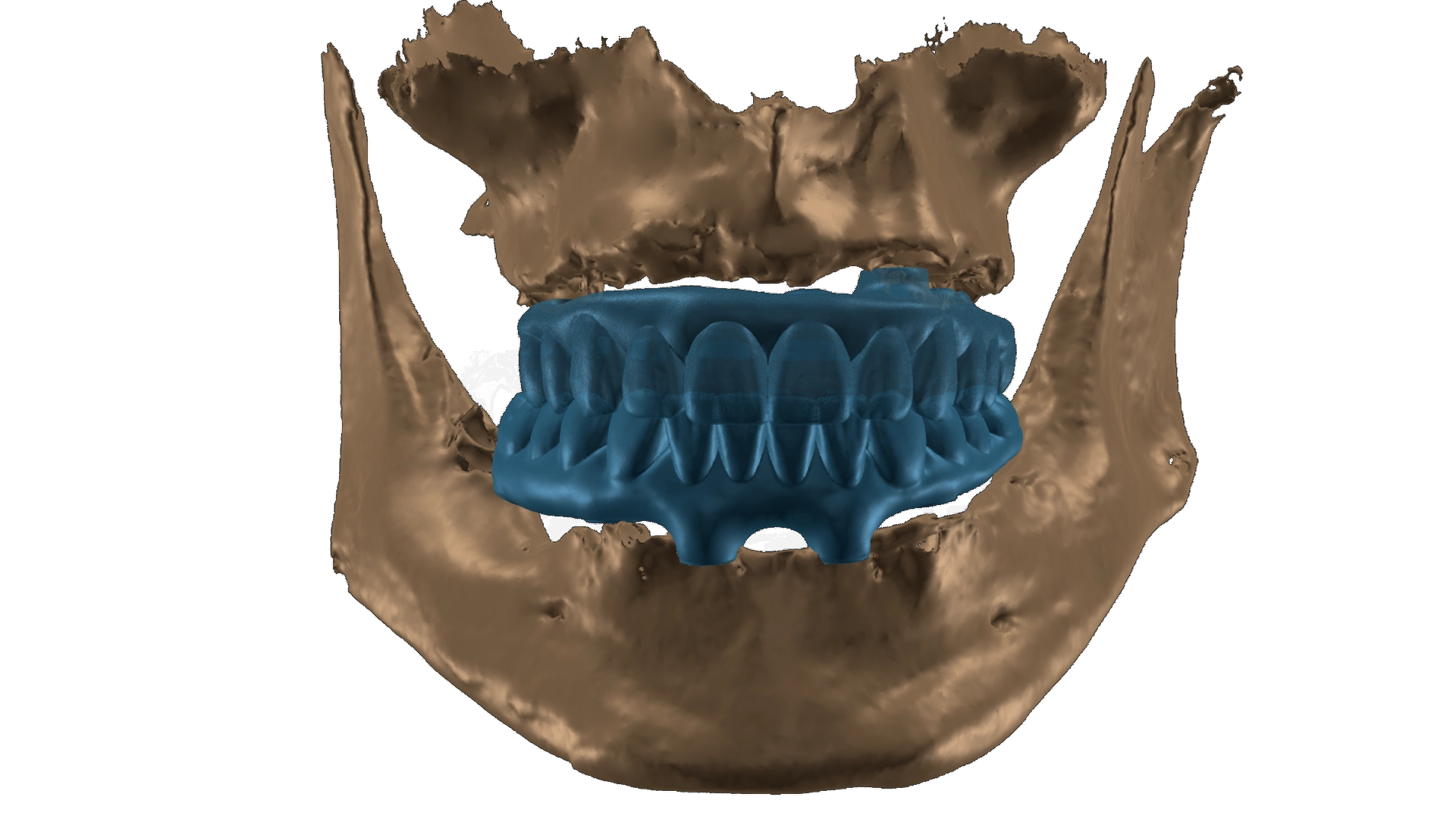
Digital implant planning is a pre-surgical protocol that models your entire procedure virtually before clinical instruments are used. Instead of evaluating bone thickness solely after making an incision, clinicians examine a complete virtual model of your jaw.
This approach treats implant placement as an interconnected medical process rather than an isolated surgical event. Understanding bone density, gum contours, and adjacent roots in advance removes guesswork from the operating room.
CBCT Scanning + Surgical Guides: The Technology Behind Precision Implants at VIDENT
The foundation of modern planning is Cone Beam Computed Tomography (CBCT). Unlike standard dental X-rays, a CBCT scan captures cross-sectional slices of your anatomy to generate a 3D rendering of bone height, bone width, and critical landmarks.
At VIDENT, this volumetric data feeds directly into digital planning software. The clinician selects the ideal implant length, diameter, and insertion trajectory relative to your bite forces.
When indicated, this digital plan is translated into a physical surgical guide. This custom device rests securely over your teeth or gums during surgery, directing surgical drills along pre-set angles and exact depths.
How VIDENT’s Digital Workflow Differs From Traditional “Freehand” Planning
In traditional “freehand” implant placement, the surgeon reviews flat radiographs, makes an incision, retracts the gum tissue, and determines the drill entry point visually based on surface landmarks.
VIDENT replaces visual estimation with an integrated digital workflow:
- Digital acquisition: A 3D CBCT scan captures your bone volume, nerve canals, and sinus boundaries.
- Virtual positioning: The ideal crown shape is designed first, and the implant is positioned beneath it to support natural chewing loads.
- Guided execution: A custom surgical guide transfers that virtual plan to the surgical site with sub-millimeter precision.
- Immediate provisional readiness: Because dimensions are determined prior to surgery, a temporary tooth can often be prepared ahead of time for suitable candidates.
Traditional Implant Planning: The 3 Hidden Risks
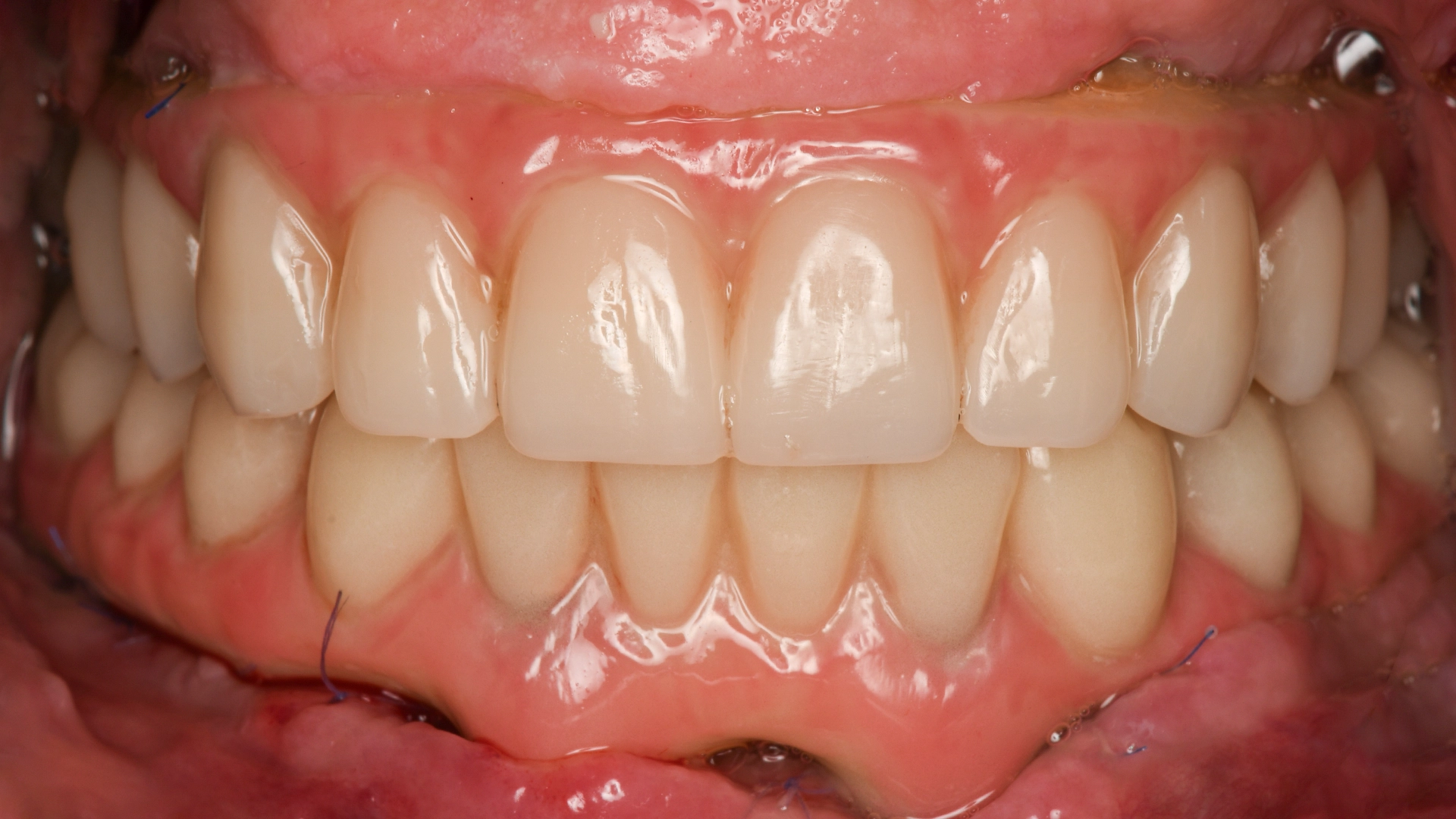
Traditional freehand techniques have restored smiles for decades. However, working without three-dimensional pre-visualization introduces distinct biological and restorative variables.
Understanding these risks helps you evaluate how modern planning protects your health and your investment.
2D X-Ray Limitations: Why Your Jawbone Looks Completely Different Than What the Surgeon Sees
A standard panoramic X-ray flattens complex three-dimensional anatomy into a single two-dimensional sheet. It reveals bone height reasonably well, but it cannot show bone width or horizontal concavities.
A jaw ridge can look deep and healthy on a 2D radiograph while actually presenting a sharp, narrow crest underneath the gum tissue. Without cross-sectional imaging, a clinician only discovers thin bone or severe undercuts after surgically exposing the bone, forcing real-time changes to the procedure.
Freehand Implant Placement and the Nerve Damage Statistics You Should Know
The lower jaw houses the inferior alveolar nerve, a major sensory pathway that provides feeling to your lower lip, chin, and teeth. In the upper jaw, the maxillary sinus floor lies directly above your molar roots.
Clinical literature shows that freehand placement carries higher rates of positional deviation compared to guided protocols. Even a minor angular discrepancy can direct an implant fixture closer to a nerve canal or sinus cavity than intended. Utilizing 3D CBCT mapping provides exact distance measurements, establishing a mandatory safety margin around sensitive structures.
The “Surprise” During Surgery That Could Have Been Predicted (And How It Affects Your Wallet)
When bone deficiencies remain hidden until surgery begins, treatment plans must adapt on the spot. A clinician may need to perform an unexpected bone graft, adjust implant dimensions, or abort placement altogether to allow preliminary healing.
These mid-procedure discoveries create clinical stress and financial uncertainty. At VIDENT, diagnosing bone architecture in advance ensures that preparatory treatments, such as bone augmentation, are identified before surgery. This upfront clarity keeps single implant care within the expected €2,500 to €3,100 range, eliminating surprise add-on fees.
Digital vs. Traditional Implant Planning: A Side-by-Side Comparison (2026 Clinical Data)
Clinical studies consistently highlight measurable differences between guided digital workflows and freehand surgery.
| Clinical Parameter | Digital 3D-Guided Planning | Traditional Freehand Planning |
|---|---|---|
| Diagnostic Foundation | 3D CBCT cross-sectional volume | 2D panoramic/periapical flat radiograph |
| Surgical Accuracy | High predictability via rigid guide | Dependent on visual estimation and manual angles |
| Anatomical Safety | Exact distance to nerves and sinuses mapped | Estimated safety buffer based on flat images |
| Provisional Restorations | Pre-fabricated same-day tooth (when indicated) | Often delayed until standard healing finishes |
| Procedure Duration | Focused, streamlined, pre-mapped | Longer chair time due to intraoperative adjustments |
Accuracy: 0.5mm vs. 2.5mm Deviation, What That Actually Means for Your Smile
In clinical comparative research, freehand implant placement frequently exhibits average coronal deviations near 1.5mm to 2.5mm and angular errors up to 7 or 8 degrees. By contrast, computer-guided surgery regularly limits positional deviation to approximately 0.5mm to 1.0mm.
In practical terms, this precision preserves the delicate papilla (the gum tissue between your teeth), prevents exposure of metal margins, and ensures chewing forces distribute evenly down the long axis of the bone.
Surgery Time: 20 Minutes vs. 90 Minutes (And Why Less Time = Less Pain)
Because key anatomical decisions are resolved during the digital planning phase, guided surgery proceeds efficiently. Placing a single straightforward implant through a planned guide can take as little as 15 to 20 minutes, compared to 60 to 90 minutes for exploratory freehand surgery.
Shorter chair time is not just convenient; it is biologically advantageous. Less tissue exposure to air, reduced flap retraction, and controlled osteotomy drilling result in significantly less tissue trauma, lowering postoperative inflammation and surgical soreness.
Recovery Speed: Back to Work Tomorrow vs. Swelling for Two Weeks
Extensive surgical incisions and prolonged flap elevation trigger the body’s natural defense cascade: substantial swelling, bruising, and discomfort that can linger for days.
With digital planning, incisions can be minimized, and bone preparation is executed with high efficiency. Many patients resume light activities and work the following day with standard over-the-counter pain relievers, avoiding the prolonged swelling often associated with exploratory surgeries.
How We Use Digital Implant Planning at VIDENT
At our Antwerp clinic, digital precision serves a simple objective: helping you regain a stable, natural smile with complete peace of mind.
Our clinical protocol follows three interconnected principles.
Planning Starts With the Final Restoration
We do not ask, “Where can we place an implant?” We ask, “Where does your new tooth need to be to ensure ideal chewing function and aesthetic balance?”
Using digital design tools, we model your final crown first. Once the ideal tooth shape, height, and emergence profile are established, we determine the exact implant position below the gum line needed to support that restoration permanently.
Bone, Soft Tissue and Anatomy Are Assessed in Three Dimensions
During your consultation, our team examines:
- Bone volume and architecture: Measuring bone height, width, and localized density.
- Proximity to anatomical landmarks: Safeguarding nerve paths and sinus cavities.
- Gingival biotype and contours: Ensuring sufficient soft tissue surrounds the final restoration.
- Bite dynamics: Balancing your natural chewing forces across all teeth.
You review these findings clearly on screen during your visit, transforming complex anatomical data into visual understanding.
The Implant Position Is Planned Around the Whole Treatment
An implant is a medical investment meant to last for years. Therefore, planning accounts for every phase of your journey:
- Surgical safety: Precision placement using custom guides.
- Same-day temporary solutions: Preparing a provisional crown in advance if your bone stability allows immediate placement.
- Biological integration: Allowing 8 to 12 weeks for osseointegration, during which your bone securely fuses with the implant surface.
- Personalized comfort: Adapting local anesthesia and tailored sedation options to your individual sensitivity and anxiety levels.
Before & After: Real VIDENT Smiles
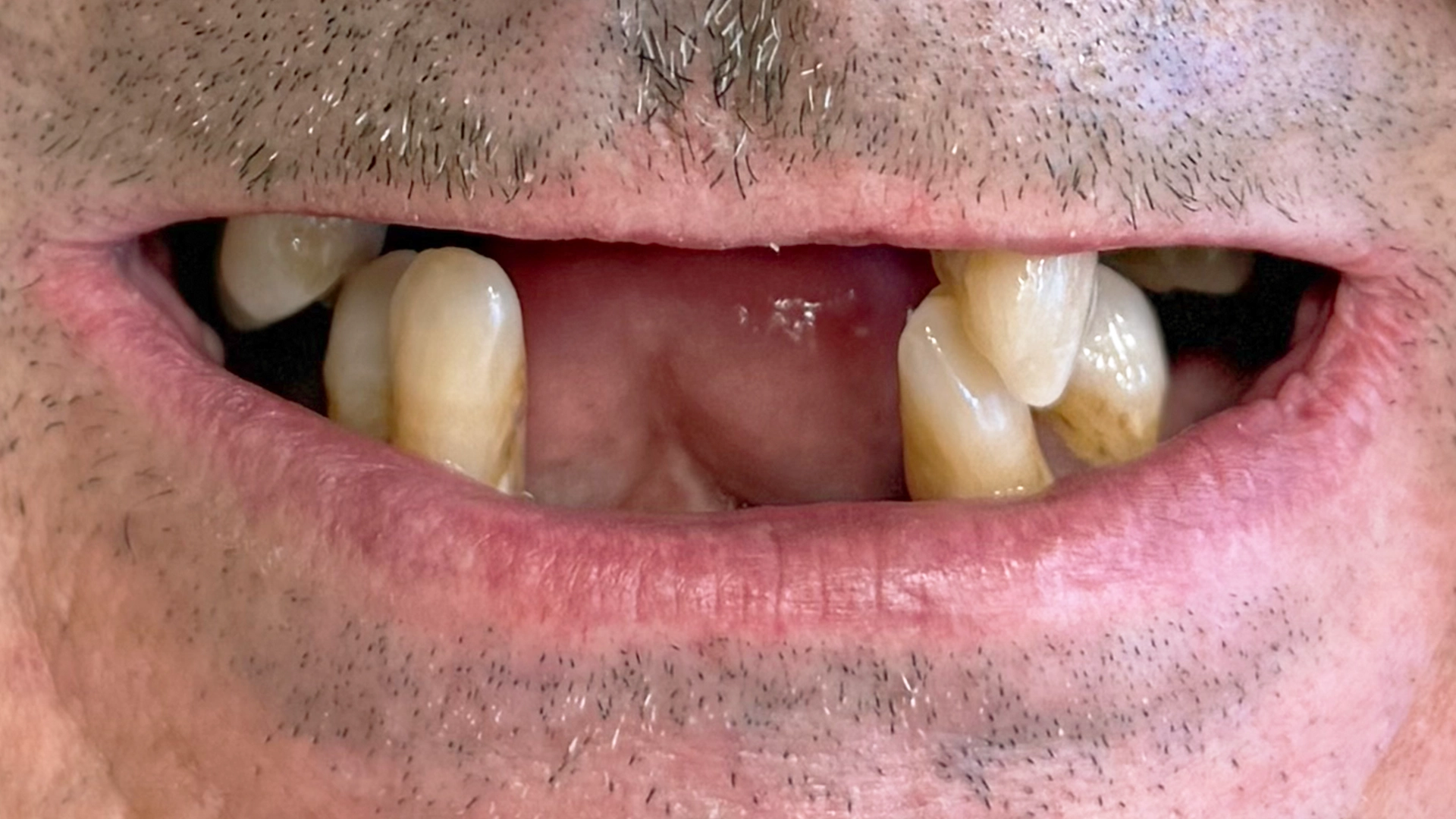
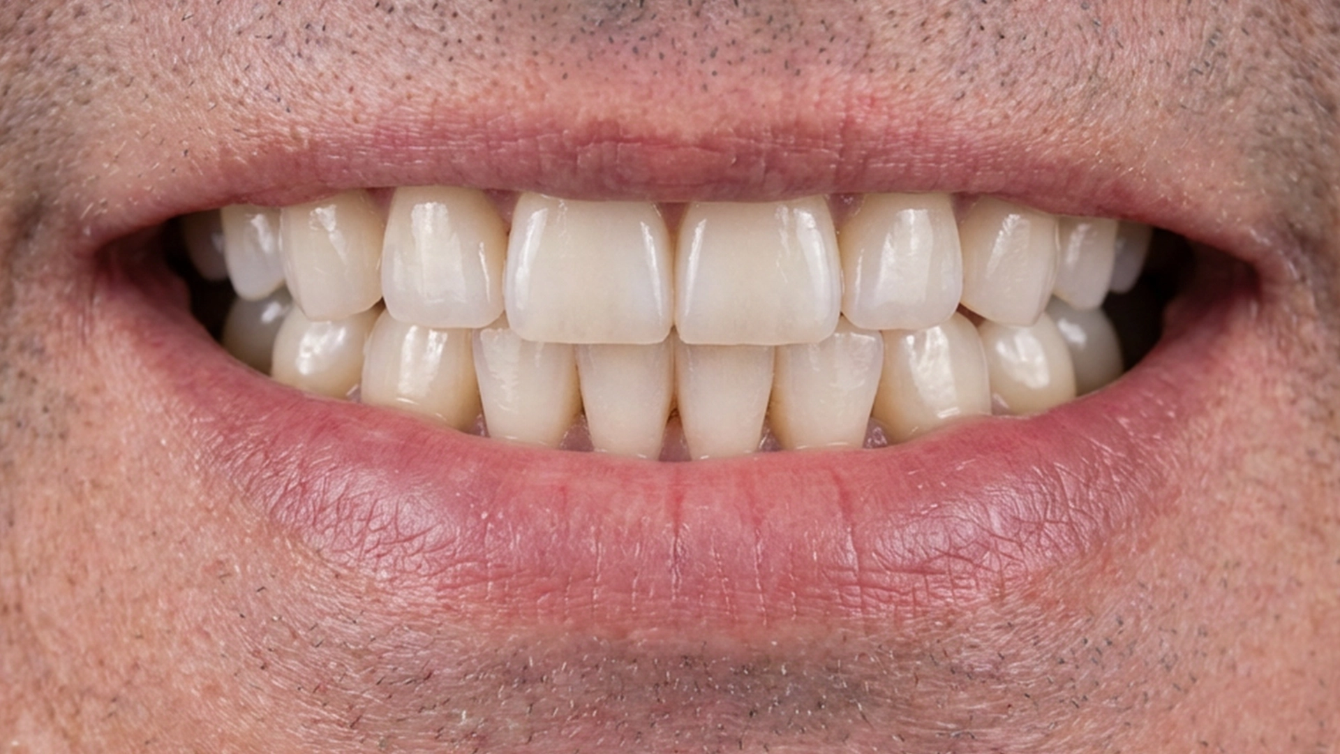
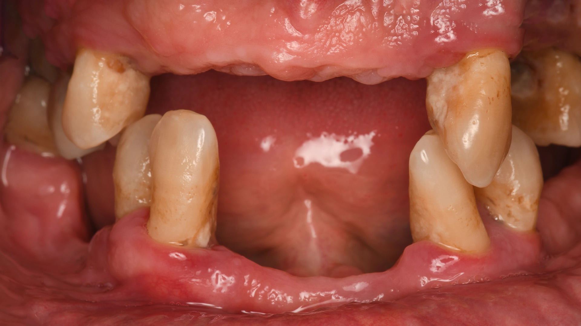
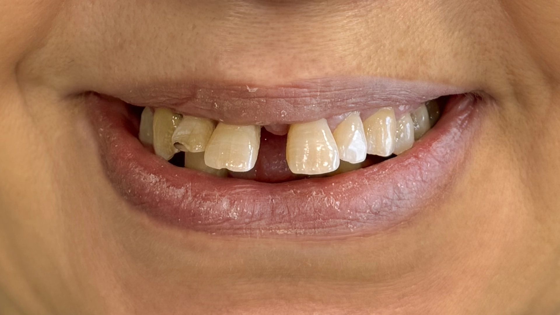
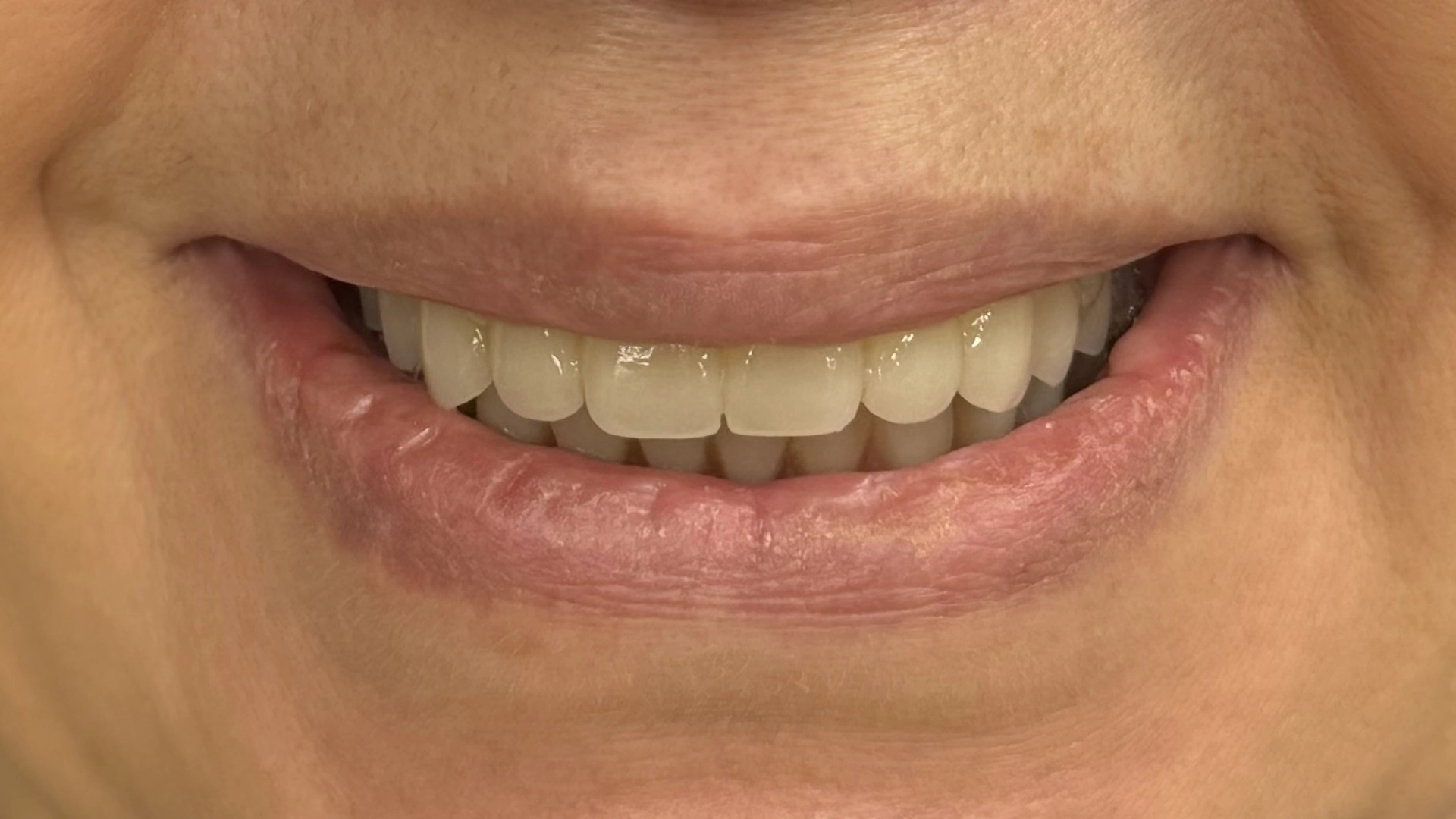
Move the slider on each case to compare the smile before treatment with the result after treatment.
 Before
 After
After
 Before
After
Before
After
 Before
 After
After
Results vary from patient to patient. Every treatment plan is based on your own anatomy and needs.
What Should You Do If You Are Considering Dental Implant Treatment?
Navigating tooth replacement does not have to feel overwhelming. When treatment is organized logically and explained with total transparency, moving forward becomes a clear, confident decision.
Start With an Assessment of Your Individual Anatomy
The only reliable way to determine if you are a candidate for dental implants is through a comprehensive clinical examination and 3D imaging assessment.
At VIDENT in Antwerp, your consultation delivers complete clarity:
- A thorough 3D CBCT evaluation of your bone and gum condition.
- An honest assessment of whether an implant, a bridge, or an alternative solution serves you best.
- A determination of whether a temporary tooth can be placed on the day of surgery.
- A written treatment plan outlining precise stages, an estimated timeline, and a transparent cost overview before treatment starts.
Frequently Asked Questions (FAQ)
Does digital 3D-guided implant surgery hurt during or after the procedure?
The placement itself is completely painless because it is performed under effective local anesthesia, with personalized sedation options available for anxious patients or complex treatments. Because 3D CBCT planning and custom surgical guides eliminate guesswork and minimize unnecessary gum incisions, tissue trauma is significantly reduced. Most patients experience only mild tenderness for 24 to 48 hours, which is easily managed with standard over-the-counter pain relievers.
Can I get a replacement tooth on the exact same day my implant is placed?
In many clinically suitable cases, yes. A same-day temporary crown or bridge can be placed immediately if your implant achieves sufficient primary mechanical stability in the jawbone and your bite forces allow safe loading. Your 3D CBCT scan at VIDENT evaluates bone density in advance to determine if immediate temporization is clinically safe for your anatomy before surgery begins.
How long does the entire dental implant process take from start to finish?
For most patients, biological healing through osseointegration—where bone cells securely fuse with the titanium surface—takes approximately 8 to 12 weeks. Once your clinician confirms solid integration, your custom-designed final ceramic crown or bridge is placed. If preparatory procedures like bone augmentation are required, the overall timeline is adjusted accordingly and clearly detailed in your written treatment plan.
What happens if my 3D scan shows I don’t have enough bone for an implant?
Bone loss is very common after tooth loss, but having reduced bone volume does not immediately disqualify you. Digital planning allows us to identify localized bone deficits with millimeter accuracy before touching a scalpel. Depending on your anatomy, we can evaluate targeted bone grafting, sinus elevation, or non-implant alternatives such as conventional dental bridges to restore your smile safely.
How much does a single dental implant cost in Antwerp, and is it covered by Belgian health insurance?
At VIDENT, a single complete implant restoration typically ranges between €2,500 and €3,100, which includes 3D assessment, surgical placement, a temporary crown (when clinically appropriate), and the custom final crown. While basic statutory Belgian health insurance (mutuelle / ziekenfonds) generally does not cover standard adult implants, supplementary dental insurance policies often provide partial reimbursement. We provide a full, itemized written estimate and paperwork to support your insurance claims.
What makes digital 3D planning safer than traditional “freehand” implant placement?
Traditional freehand placement relies on 2D X-rays and visual estimation, which cannot reveal horizontal bone concavities or the exact 3D pathway of the inferior alveolar nerve. Digital 3D CBCT planning maps every anatomical boundary beforehand. Using a 3D-printed surgical guide restricts drill trajectory and depth to sub-millimeter precision, virtually eliminating the risk of nerve impingement or sinus floor perforation.
How long do dental implants last, and how do I care for them?
With proper oral hygiene and regular preventive check-ups, dental implants are designed to last for decades and often a lifetime. Because the restoration is planned around your natural chewing forces, mechanical wear is minimized. Caring for your implant is identical to caring for natural teeth: brush twice daily, clean interdental areas with floss or interdental brushes, and attend scheduled clinical maintenance visits.
I live outside central Antwerp. Can I visit VIDENT for digital implant treatment?
Yes, absolutely. While our modern clinic is conveniently located in Antwerp (Antwerpen), a substantial portion of our implant patients travel to us from surrounding districts, suburbs, and neighboring municipalities. We routinely welcome patients from Berchem, Borgerhout, Deurne, Wilrijk, and Merksem, as well as the wider Antwerp province including Brasschaat, Schoten, Mortsel, and Edegem. Because our comprehensive digital protocol streamlines visits, combining 3D scanning, clinical assessment, and virtual treatment simulation in one appointment, patients traveling from across the Antwerp region find the process efficient, predictable, and easily manageable.